Introduction of Flagella
Flagella are the complex filamentous cytoplasmic structure protruding through cell wall. These are unbranched, long, thread like structures, mostly composed of the protein flagellin, intricately embedded in the cell envelope. They are about 12-30 nm in diameter and 5-16 µm in length. They are responsible for the bacterial motility. Motility plays an important role in survival and the ability of certain bacteria to cause disease.
Types and Examples of Flagella
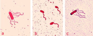
There are 4 types of flagellar distribution on bacteria:

1. Monotrichous
– Single polar flagellum
– Example: Vibrio cholerae
2. Amphitrichous
– Single flagellum on both sides
– Example: Alkaligens faecalis
3. Lophotrichous
– Tufts of flagella at one or both sides
– Example: Spirillum
4. Peritrichous
– Numerous falgella all over the bacterial body
– Example: Salmonella Typhi
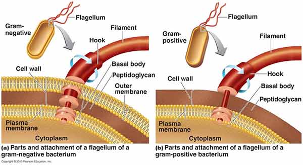
Parts of Flagella
Each flagellum consists of three distinct parts- Filament, Hook and Basal Body.
The filament lies external to the cell.
Hook is embedded in the cell envelope.
Basal Body is attached to the cytoplasmic membrane by ring-like structures.
Functions of Flagella
- Movements
- Sensation
- Signal transduction
- Adhesion
- For cells anchored in a tissue, like the epithelial cells lining our air passages, this moves liquid over the surface of the cell (e.g., driving particle-laden mucus toward the throat).
- Flagella are generally accepted as being important virulence factors
Principle of Flagella Staining
A wet mount technique for staining bacterial flagella is simple and is useful when the number and arrangement of flagella are critical in identifying species of motile bacteria.
Procedure of Flagella Staining
- Grow the organisms to be stained at room temperature on blood agar for 16 to 24 hours.
- Add a small drop of water to a microscope slide.
- Dip a sterile inoculating loop into sterile water
- Touch the loopful of water to the colony margin briefly (this allows motile cells to swim into the droplet of water).
- Touch the loopful of motile cells to the drop of water on the slide.
- Cover the faintly turbid drop of water on the slide with a cover slip. A proper wet mount has barely enough liquid to fill the space under a cover slip. Small air spaces around the edge are preferable.
- Examine the slide immediately under 40x for motile cells.
- If motile cells are seen, leave the slide at room temperature for 5 to 10 minutes.
- Apply 2 drops of RYU flagella stain gently on the edge of the cover slip. The stain will flow by capillary action and mix with the cell suspension.
- After 5 to 10 minutes at room temperature, examine the cells for flagella.
- Cells with flagella may be observed at 100x.
Staining
Observe the slide and note the following:
- Presence or absence of flagella
- Number of flagella per cell
- Location of flagella per cell
Could you please tell me why monotrichous swim faster? Is it because of their chemotropism?
These flagellum act independently on their own. So it will be more easy and simple to coordinate one flagellum(monotrichous) than anyother type.
Just like imagining running with 10 legs compared to running with two legs.
You will need more coordination to run with 10 legs.
And also it will be easy to supply enough proton motive force to power one flagellum than many flagella
Could you kindly list all possible flagellated bacteria which could cause a postive IgG or IgM 41 antibody response? Thank you!
Hello sir.. kindly tell the example for amphilophotrichous flagella
may d sky be ur limit …d diff b/w gram positive and negative are
*gram positive posses lipoteichoic acid while negative posses brain lipoprotein
*gram+ have 3 layers while gram – has 2 layers
can you please tell me the gene name which responsible for the formation of monotrichous, lophotricous, peritrichous and amphitrichous?
Nice basic information but it is not clearly mentioned the difference of Gram Positive and Gram Negative bacterial Flagellum or Flagella.
in gram +ve bacteria ,2 flagella rings are present in basal body while in gram -ve bacteria ,4 flagella rings are present