The main purpose of capsule stain is to distinguish capsular material from the bacterial cell. A capsule is a gelatinous outer layer secreted by bacterial cell and that surrounds and adheres to the cell wall. Most capsules are composed of polysaccharides, but some are composed of polypeptides. The capsule differs from the slime layer that most bacterial cells produce in that it is a thick, detectable, discrete layer outside the cell wall. The capsule stain employs an acidic stain and a basic stain to detect capsule production.
Principle of Capsule Staining
Capsules stain very poorly with reagents used in simple staining and a capsule stain can be, depending on the method, a misnomer because the capsule may or may not be stained.
Negative staining methods contrast a translucent, darker colored, background with stained cells but an unstained capsule. The background is formed with india ink or nigrosin or congo red. India ink is difficult to obtain nowadays; however, nigrosin is easily acquired.
A positive capsule stain requires a mordant that precipitates the capsule. By counterstaining with dyes like crystal violet or methylene blue, bacterial cell wall takes up the dye. Capsules appear colorless with stained cells against dark background.
Capsules are fragile and can be diminished, desiccated, distorted, or destroyed by heating. A drop of serum can be used during smearing to enhance the size of the capsule and make it more easily observed with a typical compound light microscope.
Reagents used for Capsule Staining
Crystal Violet (1%)
Crystal Violet (85% dye content) = 1 gm
Distilled Water = 100 ml
Nigrosin
Nigrosine, water soluble = 10 gm
Distilled Water = 100 ml
Procedure of Capsule Staining
- Place a small drop of a negative stain (India Ink, Congo Red, Nigrosin, or Eosin) on the slide.
Congo Red is easier to see, but it does not work well with some strains. India Ink generally works, but it has tiny particles that display Brownian motion that must be differentiated from your bacteria. Nigrosin may need to be kept very thin or diluted. - Using sterile technique, add a loopful of bacterial culture to slide, smearing it in the dye.
- Use the other slide to drag the ink-cell mixture into a thin film along the first slide and let stand for 5-7 minutes.
- Allow to air dry (do not heat fix).
- Flood the smear with crystal violet stain (this will stain the cells but not the capsules) for about 1 minutes. Drain the crystal violet by tilting the slide at a 45 degree angle and let stain run off until it air dries .
- Examine the smear microscopically (100X) for the presence of encapsulated cells as indicated by clear zones surrounding the cells.
Result of Capsule Staining
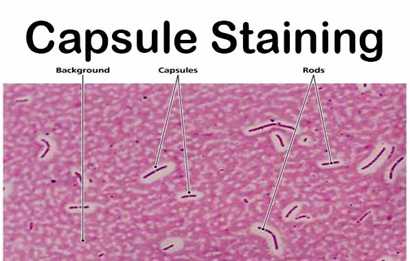
Capsule: Clear halos zone against dark background
No Capsule: No Clear halos zone
Examples of Capsule Positive and Negative
Positive
Bacillus anthracis, Klebsiella pneumoniae, Streptococcus pneumonia Neisseria meningitidis Clostridium spp, Rhizaobium spp, etc.
Negative
Neisseria gonorrhoreae, etc.
Mneomonics to remember capsulated bacteria- Some Killers Have Pretty Nice Capsule
Streptococcus pneumoniae
Klebsiella pneumoniae
Haemophilus influenzae
Pseudomonas aeruginosa
Neisseria meningitidis
Cryptococcus neoformans
Quality control of Capsule Staining
Positive control: Klebsiella pneumoniae (ATCC e13883)
Negative control: Alacilgenes denitrificans (ATCC 15173)
References
- Austin Community College
- Collin County Community College District
- Portland Community College
- Community College of Baltimore County
- Clark College
- Shah PK, et al. Practical Microbiology: Capsule Staning
- ASM Microbe Library: Capsule Stain Protocols
- Your Article Library
- HiMedia Technical Data: Capsule Stains-Kit
- Hardy Diagnostics
- Microbe Online
- Wikipedia
I have Pseudomonas aeruginosa from infected hardware. A fusion at C6-C7 level
What can anyone tell me about this type infection. What long term effects does it have to your spine or anywhere else in the body. Or long term use of antibiotics
I have been sick and have loss of motor functions
Please help me to know what’s going on